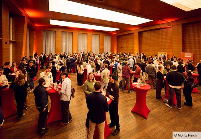
Modern methods in artificial intelligence (AI) are becoming increasingly important in science. As part of Goethe University Frankfurt’s “citizens’ university” activities, offering insights into scientific practice, Professor Eike Nagel recently showed how researchers at the Cardio-Pulmonary Institute (CPI) excellence cluster use AI in cardiac imaging. Nagel heads the Institute for Experimental and Translational Cardiovascular Imaging at Goethe University’s Faculty of Medicine, and works on developing enhanced options for the treatment of cardiovascular diseases. In an effort to make his research both more accessible and more comprehensible to everyone, Prof. Nagel opened his institute to visitors on May 10.
The evening kicked off with an introduction, as Eike Nagel explained his research focus to visitors in laypeople’s terms. Several years ago, he and his working group published a much noted study showing that a non-invasive magnetic resonance imaging (MRI) examination in the diagnostic investigation of cardiac diseases is just as efficient as classical cardiac catheterization. During this operative procedure, a fine tube is inserted into the heart via a blood vessel – an invasive means of surgery that can be avoided by performing MRI. As part of his current research, Nagel uses MRI to investigate the effects SARS CoV infections, and long COVID in particular, have on the cardiac muscle. In his presentation, Nagel also described in detail the long path from an initial observation and theory all the way to the final publication, which often requires a lot of time, patience and, not least, sufficient research funding. The lecture was followed by four fascinating “research stations” that Nagel and his eight team members had prepared to offer practical insights into the day-to-day work of a cardiovascular scientist. The practical insights underscored what he had already stressed in his initial presentation: “a good team is essential for my work!” The outstanding commitment and motivation of his interdisciplinary team, which includes medical doctors, software engineers and computer scientists, impressed participants at each of the stations.
Guessing games with MRI images
A special highlight was the MRI station. Radiology technician Tilman Ross demonstrated the machine’s capabilities using bananas, avocados, apples and kiwifruit – meaning only fruit, and no people, were exposed to its strong magnetic field. Having hid some of the fruit in a cardboard box, Ross invited onlookers to guess its contents based on the MRI images. During this exercise, it quickly became clear that different sections produce very different results – as is the case during an examination of the heart. Using a paperclip fastened to a lanyard, Ross impressively demonstrated the magnetic field’s strength: As if by magic, the paperclip was pulled toward the MRI from quite a distance. Participants had a lot of questions, many of which centered on the special safety measures necessary for an MRI examination owing to the magnetic field. Generally speaking, Ross explained, cardiac pacemakers, implants (except cochlear implants) and endoprostheses do not exclude patients from an MRI examination, adding, however, that his own experience has taught him that it’s not a good idea to bring a smartwatch close to the MRI device – the sensitive electronics are frequently destroyed.
A cardiac MRI quickly produces hundreds of images – and the doctor analyzing them not only has to do so carefully. The task is both time-consuming and requires great concentration. This is precisely where AI can be brought in for support. At the next station, software developer Simon Bohlender and medical student Quang Anh LeHongAn presented a program the team developed specifically for cardiac imaging. It evaluates the MRI images automatically, thereby relieving the doctor’s workload and reducing the time it takes to deliver a diagnosis. Ideally, the software should be even more accurate than a doctor; to that end, it has been trained with thousands of MRI images so that it can recognize and mark certain patterns and characteristics in the human heart – a process known as machine learning, which in turn is a field within AI. At this station visitors had the opportunity to compete against the AI. They had to trace on a PC the contours of the cardiac muscle in ten MRI images of a heart – and lost hands down to the computer, which was at least 50 times faster than its human competitors. All the same, Bohlender was able to allay fears that AI might replace the physician, emphasizing that every evaluation naturally has to be reviewed by a doctor. What the automated, AI-aided analysis of MRI images can do, is to considerably shorten the time to diagnosis.
Using AI to sift through the research literature
There are other areas, too, where science has become practically inconceivable without AI. At the next station, Deniz Desik and Paul Weibert from Nagel’s team spoke about the constantly rising number of scientific publications. In 2021 alone, a new publication on the topic of COVID appeared every two minutes, illustrating the fact that it is becoming increasingly difficult for scientists to gain an overview of the enormous quantity of relevant publications. Portals such as Semantic Scholar already use AI to understand the semantics of scientific literature and to support scientists in their search for relevant research. This was certainly the most challenging of the four stations for laypersons; after all, the code presented, which is used to search for publications, seems very complicated to non-IT experts at first glance. Thanks to Deniz and Paul, however, it quickly became clear that a search using fewer keywords at the right place is actually not that difficult.
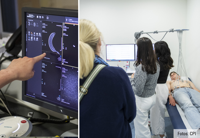
AI is already deployed outside laboratories and hospitals, in very ordinary situations and objects we use every day, like smartphones and smartwatches. At the fourth station, medical technician Aydan Özkan and doctoral student Laura Pappas demonstrated that fact very interestingly using an electrocardiogram (ECG) – an examination virtually everyone is familiar with. An ECG is especially important in the diagnostic work-up of cardiac arrhythmias and heart attacks. To start with, Özkan and Pappas performed a classical 12-channel ECG on a volunteer and explained its evaluation, vividly illustrating how the ECG machine makes a computer-aided evaluation, and can recognize a healthy sinus rhythm or a dangerous atrial fibrillation during the procedure. Things became particularly exciting when the team then recorded an ECG on an Apple-Watch. To do so, all it takes is to touch the sensor of the watch (on one’s wrist) with a finger of the other hand and start the recording. Although the 1-channel ECG integrated into this smartwatch cannot recognize every type of heart disease, it is capable of indicating cardiac arrhythmias such as atrial fibrillation very reliably and is therefore definitely a useful option for long-term monitoring, especially for high-risk patients.
“It was a great event – complete with a nice team offering fascinating information. I would love to come back another time!” said one participant as the evening ended. Nagel’s team succeeded in giving all visitors a fascinating peek into cardiovascular research and demonstrating the areas in which AI can be deployed to benefit the patients.
Claudia Koch & Daniela Daume


