How traditional EEG is experiencing a renaissance in stroke therapy
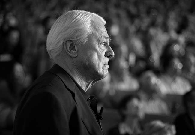
It’s an exciting time for clinical neurophysiology. New scientific and technical advances are making it easier to study motor networks in the brain and – since recently – use non-invasive procedures to treat them. To customize therapies for stroke patients, neurologist Christian Grefkes-Hermann from Frankfurt University Hospital combines electroencephalography (EEG) – a 100-year-old method for measuring brain activity – with magnetic fields that stimulate the brain.

Let’s start with the good news: At 15 percent, the mortality rate of stroke patients has almost halved over the past 25 years. This is thanks to improvements in the acute care afforded by stroke units. When a vascular occlusion or, less frequently, a cerebral hemorrhage disrupts blood flow in the brain, it may cause functional deficits such as paralysis as well as speech, vision or other impairments that “strike” a person out of the blue. If a vascular occlusion is not remedied within a few hours, normal functions are permanently lost and in the worst case the patient can die as a result.
In line with the maxim “Time is Brain”, i.e. time is of essence when it comes to stroke treatment, effective procedures have been developed to restore blood flow in stroke-affected brain regions, which are being implemented in clinical care for many years. For example, blood clots are dissolved by administering appropriate drugs or surgically removed with a catheter. As a result, almost a third of all stroke patients are left with no motor or speech impairments. As a positive side effect, neurology has developed from a medical specialty into recognized “emergency care”.
The bad news is that strokes are still the most common cause of acquired disability in Germany, where every year around 270,000 people suffer a cerebral infarction. In view of the demographic trend, this figure can be expected to rise in the coming decades. Thus, although acute care saves lives, it cannot prevent over 50 percent of all patients from experiencing moderate to severe impairments afterwards. And even worse: Many stroke patients who have undergone rehabilitative treatment following the acute post-stroke phase are often left with no further treatment despite still being partly paralyzed or experiencing coordination problems. “We assume that there are currently around one million stroke survivors,” says Professor Christian Grefkes-Hermann, 47, describing the status quo. The neurologist highlights that medicine often “loses sight of these people” after acute stroke care and rehabilitation – a problem that must urgently change.
Individual treatment
Grefkes-Hermann has specialized in the motor networks of the brain and the rehabilitation of motor dysfunction after brain damage. He has been the director of the Neurology Department at University Hospital Frankfurt’s Center for Neurology and Neurosurgery since 2023. His aim is to offer therapies that improve functional recovery for patients who have experienced a stroke or are suffering from other neurological disorders such as Parkinson’s disease or inflammation of the brain. More precisely: In collaboration with colleagues from neighboring disciplines such as neurosurgery, neuroradiology, vascular surgery and cardiology, stroke patients in Frankfurt should be given access to individualized treatment – as is already increasingly the case with cancer or heart disease.
Part of the groundwork for developing Frankfurt into Germany’s top hub for neurology was laid just recently: The Neurology Department has been given a new, bigger building on the grounds of University Hospital Frankfurt, which has provided space for a larger stroke unit for the acute care of patients and the planned Brain Stimulation Outpatient Clinic for the further therapeutic treatment of stroke patients.
Dynamic network
There are further reasons for believing that Grefkes-Hermann will be able to make significant progress in the development of non-invasive neurophysiological diagnoses and therapies for people with brain injuries in the near future: Over the past 15 years, technical and scientific advances have seen to a considerable growth in medical knowledge and brain mapping. “Even if we do not yet fully understand all the brain’s functions,” as Grefkes-Hermann says, “it is clear that the human brain, which controls unique and complex phenomena such as language, memory, emotions and motor skills, must be understood as a dynamically functional, highly complex network: Its cognitive functions can less be attributed to individual specialized regions, but rather build on the networking and interaction of various such regions.”
During his professional career prior to joining Goethe University Frankfurt, Grefkes-Hermann gathered substantial knowledge of how brain regions interact, but also of how functional brain networks regenerate and reorganize themselves, for example in neighboring regions or by integrating influences from more distant brain areas. As a senior consultant and Professor for Stroke Recovery and Neurorehabilitation at University Hospital Cologne as well as the head of a research group, first at the Max Planck Institute for Neurological Research (now the Max Planck Institute for Metabolism Research) in Cologne and then at the Institute of Neuroscience and Medicine at Forschungszentrum Jülich, he has been working for many years on brain networks, the mechanisms of functional recovery and the effects of brain stimulation. He also transferred ongoing research projects funded by the German Research Foundation (DFG) with him to Frankfurt.
Renaissance of the EEG

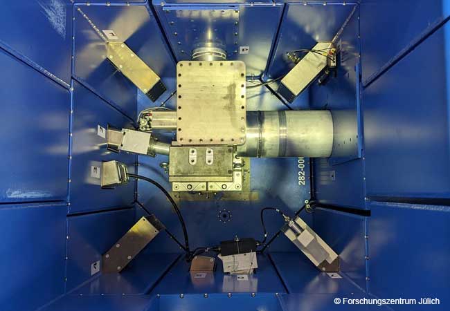
In order to reveal the key mechanisms underlying human brain function, the neurophysiologist and his team have contributed to the renaissance of a non-invasive procedure: electroencephalography (EEG). After its discovery almost exactly 100 years ago, this method for measuring brain activity was primarily used in the diagnosis of epilepsy and reduced states of consciousness, but since the 1990s and 2000s it has been adopted more and more to analyze brain function after stroke. The EEG was somewhat overshadowed by the introduction of new imaging techniques with better spatial resolution, such as functional magnetic resonance imaging (fMRI). The principle behind electroencephalography is that nerve cells generate electrical fields that are measurable outside the brain. These measurements are conducted via electrodes attached to the scalp. Time series from the individual electrodes deliver information about brain functions in real time. While the EEG, which has meanwhile become more technically advanced owing to more sensitive sensors and better amplifiers, provides increasingly differentiated insights into the temporal domain of neuronal activity in the range of milliseconds, the fMRI technique is still fascinating because of its high spatial resolution with millimeter precision and better spatial resolution.
Grefkes-Hermann is now utilizing the advantages offered by these techniques for both understanding human brain function as well as developing new treatment approaches: on the one hand, to assess temporal sequences of brain activity and, on the other, to produce spatial images of this activity. For diagnosis, Grefkes-Hermann mostly uses electroencephalography – not least because it is “far cheaper and more feasible than MRI” for broad clinical use. Indeed, measuring brain activity reveals abnormal changes in nerve cells and network nodes in stroke patients, making it possible to predict which patient will recover from their stroke-induced neurological deficits. An EEG can predict, for example, whether and to what extent certain brain regions are treatable or will regenerate.
Help from artificial intelligence
Grefkes-Hermann’s non-invasive treatment approach works with strong magnetic fields. The method he uses is called transcranial magnetic stimulation (TMS) and was first presented in 1985 by Anthony Barker, an English scientist and researcher at the University of Sheffield. TMS is applied by means of copper coil positioned on the scalp that generates a strong magnetic field for around 50 microseconds, triggering an electrical field in the cerebral cortex. The energy impulse triggers a cascade of neuronal potentials, which prompt communication between the cells in connected brain regions.
The close coordination of EEG and TMS in this therapeutic procedure is also thanks to artificial intelligence (AI). Firstly, through data compression, AI recognizes patterns in neuronal activity and network configurations in rich data sets evoked by internal or external stimuli. Secondly, AI may predict the temporal profile of individual EEG rhythms, allowing for very precise stimulation of particular brain states known to enhance or suppress cortical excitability. “If you stimulate nerve cells with magnetic fields in a specific rhythm,” explains Grefkes-Hermann, “they can be activated or dampened.”
Various causes
Christian Grefkes-Hermann’s research has contributed to furthering our understanding of how motor network changes relate to the recovery of functions. He explains that the brain already starts to repair itself in the first few hours after a stroke. “But the brain can also take a wrong turn along the way.” In other words, certain brain regions sometimes respond so over-actively during regeneration that they may interfere with the coordinated activity of others. In other regions, in turn, the nerve cells are no longer active at all. Importantly, stimulation of a particular region may activate the entire network of interconnected areas, thereby having a brain-wide treatment effect supporting the reorganization process to overcome the loss of functions.
But why do patients react differently to treatment? Neuroimaging and EEG assessments have shown that similar neurological symptoms after a stroke can be caused by very different lesions. “The reorganization pathways in the brain can be very different between individual patients. Each region is connected in a unique way and responds differently to a lesion,” says Grefkes-Hermann. “This explains why there is no one-fits-all therapy for all patients. We need to decipher the individual network malfunction caused by a specific lesion in order to offer a targeted treatment.” It is, however, not just a question of when and where brain regions have been damaged: Age, gender and other factors also influence the brain’s ability to regenerate.
Stimulating the brain
For all patients, however, the general therapeutic rule applies “the earlier the better”. Grefkes-Hermann emphasizes: “We have good evidence that starting rehabilitative treatment already in the first week after stroke leads to significantly better outcomes.” This also applies to brain stimulation treatments activating the motor regions of the lesioned hemisphere, allowing for better reorganization of brain networks. What is surprising, says Grefkes-Hermann, is that when the hand region is stimulated, some patients also report improvements of mouth control, probably caused by stimulation of neural connections between the respective brain regions. In this way, stroke therapy is delivering insights that in turn feed into basic research of the human brain.
In therapeutic practice, magnetic brain stimulation proves particularly effective when the procedure is performed immediately prior to physiotherapy and occupational therapy. This is another reason why Grefkes-Hermann is working on the implementation of this technology in rehabilitation centers as soon as possible. “There is a tremendous need to supplement traditional rehabilitative interventions with innovative brain stimulation,” he says. A prerequisite for this is that the medical professions involved in treating patients – nurses, doctors and therapists – are trained in these new methods. “To date, the use of non-invasive therapies in clinical practice is not always state-of-the-art,” says Grefkes-Hermann. “Although there are international recommendations on how magnetic stimulation should be applied, they are not always implemented.” That is why he is also forging ahead with appropriate training, for example by developing a curriculum for the training academy of the German Society for Clinical Neurophysiology and Functional Imaging (DGKN). The TMS Outpatient Clinic in Frankfurt, which just opened in 2025, will also be such a place for further training. Christian Grefkes-Hermann, his team and colleagues from other disciplines at University Hospital Frankfurt are excited. “It is amazing to see that patients can get better with TMS treatment, helping them to better master their everyday life, ultimately leading to better social participation and quality of life. And for us, restoring that,” says Grefkes-Hermann, “is what it’s all about.”

About / Christian Grefkes-Hermann, born in 1977, studied medicine in Düsseldorf, Sydney and London, earned his doctoral degree at Heinrich Heine University Düsseldorf in 2005 and his postdoctoral degree (Habilitation) at the University of Cologne in 2011. In 2013, he was appointed as Professor for Stroke Recovery and Neurorehabilitation at the University of Cologne, which also involved responsibility for the working group “Rehabilitation of Cognitive Disorders” at Forschungszentrum Jülich. In 2023, he accepted a call from Goethe University Frankfurt and has since been the director of the Neurology Department at University Hospital Frankfurt. Grefkes-Hermann is past-president of the German Society for Clinical Neurophysiology and Functional Imaging (DGKN) and spokesperson for the Interdisciplinary Neurovascular Network in the Rhine-Main region (INVN Rhine-Main).
grefkesh@uni-frankfurt.de

The author / Pia Barth studied philosophy and literature and works as a science communication officer and public relations editor at Goethe University Frankfurt.
p.barth@em.uni-frankfurt.de