Goethe University Frankfurt is applying for the upcoming round of the Excellence Strategy of the German federal and state governments with four new clusters on the following research topics: Trust in conflict (ConTrust), infection and inflammation (EMTHERA), the origin of heavy chemical elements (ELEMENTS), and cellular architectures (SCALE). These applications bring together the competencies and pioneering ideas of Goethe University Frankfurt with those of colleagues in the Rhine-Main Universities (RMU) alliance and additional partners from four major non-university research organizations. The Cluster of Excellence “Cardio-Pulmonary Institute” (CPI), first set up in 2019, will submit a full proposal next year.
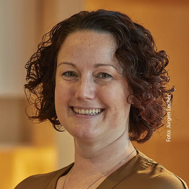
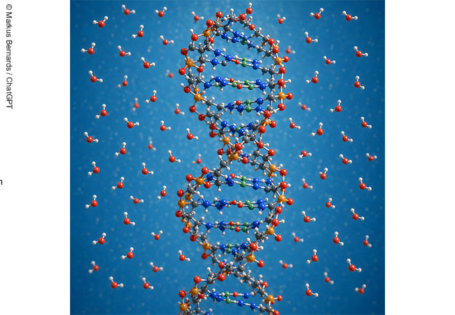
Bonnie Murphy conducts research on what is arguably the most versatile group of cellular building blocks: proteins. To be more precise, the working group leader at the Max Planck Institute of Biophysics is passionate about molecular machines, i.e. complexes of proteins that work together to accomplish a specific task, such as build something, utilize food, or protect the cell. „I want to understand how these machines work, and am particularly interested in how their structure affects their function,“ Murphy explains. Her team specializes in the use of cryo-electron microscopy (cryo-EM), which can reveal details all the way down to almost the individual atom. Rapid freezing (kryos = Greek for cold) preserves the samples in their respective state before microscopy. Using image-processing software to detect small differences in these snapshots it then becomes possible to reconstruct the manner in which the molecular machines move – „and thus how they could possibly function,“ the structural biologist explains. Electron irradiation produces an image of the samples – or, to be more exact, a map of their electron density. By combining many images, this two-dimensional black-and-white image can be used to reconstruct a three-dimensional image, which, in turn, can be converted into an atomic model. To do this, it is helpful to have information about proteins in advance, Murphy explains: „Proteins consist of a chain of amino acids that is folded in a more or less complex manner. The sequence of amino acids makes it easier for us to determine what must be in which position in the atomic model.“
From molecule to cell
To understand how an entire cell functions, however, takes more than just understanding how individual molecular machines work. „The cellular organization of the cell also plays a crucial role,“ Murphy says. This cellular architecture is at the heart of the new SCALE (Subcellular Architecture of Life) research network, which is currently applying for funding from the German Research Foundation as part of the Excellence Initiative. „The focus in SCALE is on finding out how its internal organization enables the cell to function as a unit,“ summarizes Murphy, who is also the working group leader, adding that cryo-EM – and cryo-tomography in particular – are ideally suited to address this question. Tomography involves the use of an electron microscope to take a large number of images from different angles, which can then be assembled into a three-dimensional image.
A cell’s internal organization – for example, there being organelles, i.e. separated compartments surrounded by a membrane – has consequences for biomolecules. As such, proteins are exposed to very different conditions depending on where in the cell they are located – in the cytoplasm, in an organelle or in an enveloping membrane: here, they come into contact with different ions, pH values or lipids that form the membranes of the cell and its compartments. „All of these factors enable the complex ‚dance‘ that allows individual molecules to cooperate in such a way as to create a functioning cell,“ Murphy says. That is why it is important to be able to image such factors with cryo-EM and understand how they vary depending on the subcellular architecture.
Detailed images
A vivid example of this is auxiliary factors that are important for activity in many molecular machines. „Because these metals, lipids or small molecules are not encoded in the amino acid sequence, finding them in the reconstruction of an electron microscopy image is particularly challenging for structural biology,“ Murphy says. That is why she and her team are working on further developing cryo-EM techniques, so these factors are better understood. Techniques used in materials science to detect elements are opening up entirely new possibilities. That being said, most inanimate materials are insensitive to electron bombardment; biomolecules are in fact destroyed by it, which is why they may only be bombarded with a few electrons. The result is that the individual images show only few details. „It is exactly for this reason that we assemble many individual images into a reconstruction,“ Murphy reveals. „By combining analytical EM techniques with cryo-EM image processing algorithms to reconstruct three-dimensional images, we prevent too much irradiation in the microscope from destroying our biological samples and still get detailed images.“ Progress already has been made in applying this technique to individual molecular machines, and Murphy now hopes „to apply this to the study of subcellular architecture within SCALE.“
One focus of SCALE is on mitochondria – the so-called „power plants of the cell“, where nutrients are broken down to produce energy. „Mitochondria are essential to the survival of every cell, and their failure is associated with a variety of diseases,“ Murphy says. „In SCALE, we want to find out how the structure of mitochondria affects their function and how that contributes to keeping the cell healthy.“ That is also where the new cryo-EM techniques will come into play.
Making living cells visible
How the spatial arrangement of proteins in the cell affects their function is also of interest to Mike Heilemann, a fellow member of the SCALE consortium who conducts „optical cell biology“ at Goethe University’s Institute of Physical and Theoretical Chemistry. „We are developing methods with which to make the spatial arrangement of proteins in cells visible,“ the chemist explains. Since a bombardment with electrons is out of the question when it comes to visualizing this in living cells, Heilemann uses light microscopy, or, more precisely, fluorescence microscopy. „For this purpose, we label biomolecules with fluorescent dyes, which glow when prompted by laser light, allowing us to see the labeled molecule in the light microscope.“
Light microscopy is perfect for examining entire cells, but the resolution does not suffice to visualize organelles or individual proteins. This distance, at which two points can just still be perceived separately, corresponds to about half the wavelength of the light used. „For light microscopy, this puts us at about 200 to 300 nanometers,“ explains Heilemann. Although a protein is usually only a few nanometers in size, using fluorescent dyes, it can still be made visible individually. If, however, proteins are densely packed together, as in a cell, the resulting image is smudged. „That is precisely what we want to change,“ the microscopy expert explains.
Neuronal networks improve resolution
Heilemann describes the trick of so-called single-molecule localization microscopy as follows: „We reprogram the fluorescent dyes in such a way that only one protein per time unit lights up in the crowd. By separating the signals in time, we can determine the position of each protein independently of the resolution limit.“ Two methods are currently used to do this: Either an additional step is incorporated that allows the fluorescent dyes to emit light in the first place, or the fluorescent probes are designed to bind only very briefly to biomolecules. As a result, only a few proteins light up at a time, and in a random distribution, allowing their position to be determined without proteins that are close to each other getting in one another’s way. However, since each protein must be looked at individually, single-molecule localization microscopy is a very slow endeavor. With the help of artificial intelligence, Heilemann’s team has been able to solve this problem, too: „Based on our microscopy images, we trained a neural network to determine the position of molecules that are very close to each other, allowing us to speed up high-resolution microscopy and also visualize fast processes in living cells, including the structural dynamics of organelles.“ Once the neural network is trained for a particular microscope, it provides a kind of digital extension that can be repeatedly used.
Exchange between disciplines
Among others, Heilemann’s lab uses this technique to research growth receptors, which play an important role in cancer. „Located in the cell membrane, the receptors sense certain messenger substances on the outside of the cell,“ Heilemann explains. „After binding a messenger substance, they transmit a signal with instructions for action to the inside of the cell. What type of signal exactly depends largely on which other proteins the receptors form complexes with. That is something we can study with our microscopes.“ Within the context of the SCALE cluster, the microscopy expert sees his role as a versatile method developer. „The idea behind SCALE is to bring together many different methods and, by integrating this data, create a kind of digital twin of the human cell.“ This digital cell is then used to simulate experiments in advance, including the effects of treatments.
The chemist is convinced that one of the main advantages of consortia like SCALE is that people from different disciplines exchange ideas with each other and develop new ones based on this knowledge exchange. His own team also consists of researchers from a wide range of disciplines. „SCALE is really at the heart of my interests thematically,“ Heilemann says, adding that while he anticipates many challenges, „that’s what makes it so exciting! If we manage to create such a digital twin, we can say we’ve truly understood the cell.“ Murphy also believes SCALE is a great framework for bringing together groups with different expertise. Since she and her team have so far worked primarily at the level of individual machines, she sees the consortium as an opportunity to learn more about more complicated systems. „We’re looking forward to being able to further develop our methods as part of the SCALE cluster, to understand how organelles and cells work.“
Larissa Tetsch
SCALE
The name of the SCALE cluster initiative stands for „Subcellular Architecture of Life.“ The research network brings together researchers from Frankfurt whose focus lies on studying the structure of the human cell.
To this end, they are using and developing sophisticated techniques to make the smallest details visible. As part of this effort, the initiators of SCALE consciously want to build on a traditional strength of researchers at Goethe University’s Riedberg Campus.
Further information here.